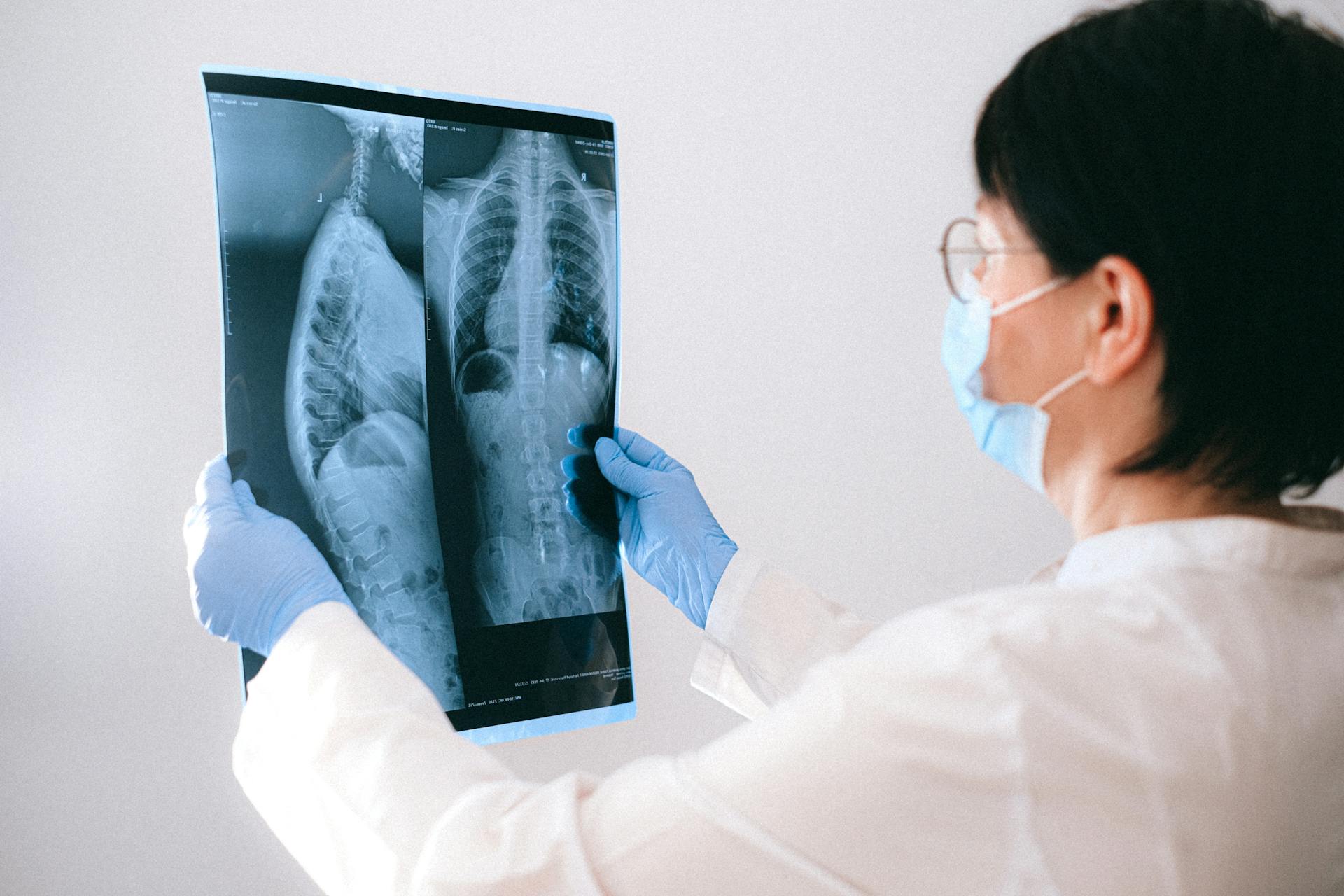
When it comes to diagnosing a potential concussion, an MRI scan can provide important information for radiologists to consider. But does an MRI alone provide enough evidence of a concussion for a diagnosis? In other words, can a radiologist diagnose a concussion from an MRI scan?
The answer is not entirely clear cut. Simply put: An MRI can provide important evidence for suspected cases of concussions, but it’s only one element and does not guarantee or rule out a diagnosis. Thus, many other factors must be taken into consideration as well. Typically, doctors look at the entire medical history of the patient—including any symptoms that may have been reported—in addition to the results obtained from an MRI scan and any relevant physical testing before reaching a concusssion diagnosis. Furthermore, while an MRI may prove helpful in detecting tissue damage (or lack thereof) associated with trauma and brain injuries that could indicate a concussion, it cannot conclusively confirm the diagnosis in all cases.
When it comes to diagnosing a potential concussion — or ruling out any kind of serious head injury — using all relevant data sources is key. While MRIs are valuable tools for detecting possible signs of trauma and can potentially play an important role in diagnosis, they should be used in combination with other sources such as extensive medical evaluations and patient surveys to provide healthcare providers with more comprehensive information on which to base their diagnoses.
Discover more: Patient Request
How is a concussion identified on a CAT scan?
Concussion is the most commonly reported form of traumatic brain injury, and it can be identified on a CAT scan. A CT (computed tomography) scan, commonly referred to as a CAT scan or computed tomography, is an imaging test that produces detailed pictures of the structure inside your head and allows your doctor to look for signs of a concussion.
When the scan reveals evidence consistent with a head injury or trauma, the radiologist interpreting the images will be able to identify signs of a concussion. Depending on what type of injuries were sustained, soft tissue swelling may be visible on a CT scan. The resulting scans can also show changes in brain volume due to edema and evidence of skull fractures resulting from blunt head trauma. Other possible indications of a concussion on imaging include subdural hematoma and diffuse axonal injury pattern typical for diffuse axonal injuries in severe brain trauma cases.
Doctor will often order an ultrasound after a CT scan if there's chance that blood may have accumulated near where the head was hit, which would indicate hematoma formation in the brain tissue due to severe force trauma. In addition to being able to identify signs of concussion on CT scans and ultrasounds, it is also important for doctors to take into account symptoms correlated with a concussion such as vomiting, headache or balance problems while attempting diagnosis as well.
For more insights, see: Trauma Related
Does a CT scan show hemorrhage associated with a concussion?
A CT scan is one of the most common diagnostic tests when it comes to investigating potential head injuries, including concussions. However, while they can tell doctors a lot about the underlying condition of the brain, CT scans are not specifically designed to show hemorrhage associated with a concussion.
Hemorrhage or bleeding means that small blood vessels in the brain have ruptured or torn, and is a serious complication associated with head trauma. To see if there is any significant amount of bleeding in the brain, doctors typically look to either an MRI or CT angiogram.
MRI scans offer much better detail than CT scans since they do not rely on radiation to generate images. They usually produce more uniform images and are capable of revealing more subtle changes within tissue structures – including evidence of blood pooling and aneurysm. In addition, it has been found that MRI is much more sensitive to areas where very small amounts of blood may have accumulated - making them more accurate for diagnosing of hemorrhage from concussions.
So while a standard CT scan can help doctors spot some things related to head trauma like skull fractures or mass lesions, if there’s reason for concern about a possible brain bleed associated with a concussion then additional imaging tests such as MRI or CT angiogram must be used before coming to any conclusions.
For more insights, see: Which Meter Would Most Likely Be Associated with a March?
Does a CT scan detect subtle traumatic brain injury after a concussion?
Traumatic brain injury (TBI) is one of the most serious injuries a person can sustain, and is caused by a jolt or blow to the head. However, it can sometimes be difficult to diagnose with standard diagnostic tests and techniques as the injury may be subtle. In such cases, a CT scan may be used to detect any damage or bruising on the brain.
A CT scan is an imaging technique which can create a detailed view of the skull and its contents. A 2D image is created from X-ray beams that are projected through the head from many angles, allowing doctors to monitor any changes over time for signs of subtle traumatic brain injury after a concussion. It is also known as computed tomographic analysis, or CAT scan for short.
CT scans have been found to detect soft tissue changes in the brain including contusions, edema and other signs of injury that are difficult to assess with even an MRI scan. Studies have shown that these scans are more reliable than traditional medical tests such as neurological tests and CT head scans since they can provide an early warning system for potential problems in cases where there may not be obvious symptoms yet.
Based on scientific evidence and studies, it is clear that a CT scan can help detect subtle traumatic brain injury in patients who have suffered concussions or similar head trauma. This type of imaging has become invaluable in helping doctors make accurate diagnoses and manage these conditions more effectively.
If this caught your attention, see: Are Doctors First Responders?
Can a CT scan determine the severity of a concussion?
Concussions are one of the most common forms of traumatic brain injury and can range from mild to severe. But can a CT scan determine the severity of a concussion?
CT scans, or computed tomography scans, are a type of imaging used to visualize the structural details inside a person's body. They're often used for diagnosing and assessing a variety of medical conditions, but their usefulness in accurately determining the severity of concussions has been somewhat controversial. Studies have shown that CT scans may not be entirely reliable sources for yielding information about concussion severity as they measure physical changes in the brain rather than functional changes. Evaluation of certain types of brain lesions—such as edema, contusions, and hematomas—may also be more sensitive when done by MRI or SPECT diagnostic imaging rather than CT scanning.
That said, it's important to note that if someone on your team is suspected to have suffered a concussion, seeking and getting an immediate professional medical evaluation through neuropsychological testing should still remain top priority. CT scanning may still have its place here as it can help rule out other potential causes and complications associated with concussions—such as skull fractures and bleeding inside the brain—which require more urgent intervention such as surgery. Ultimately, it'll be up to your doctor to decidethe best combination of tests necessary for an accurate diagnosis and evaluate treatment options alongside you and your loved ones.
Explore further: Would You Rather Youtubers?
What can be seen on a CT scan that indicates a concussion?
A CT (computerized tomography) scan is a highly accurate tool for detecting issues or abnormalities in the brain. A CT scan is often used to aid in diagnosing a concussion, which occurs when there is trauma to the head that causes the soft tissue of the brain to be damaged. On a CT scan that indicates a concussion, radiologists may notice swelling and abnormal tissue changes throughout the brain, which can include bleeding or white matter abnormalities.
The presence of an increased number of “swirls” on the CT scan can indicate areas where shearing forces have caused tears in intracranial tissues, leading to a concussion diagnosis. Diffuse axonal injury (DAI), which is another form of traumatic brain injury, can also be identified through a CT scan with indications such as stretching and shearing of axons throughout several regions of the brain. Other cranial nerve injuries commonly found after suffering from a concussion can also be identified through a CT scan, such as facial nerve injuries or cochlear nerve injuries.
Finally, changes in intracranial pressure can be seen on an image taken from a CT scan and can be an indicator of post-concussion syndrome due to elevated pressure within the cranium due to compromised pathways for cerebrospinal fluid drainage within these concussive injuries. With these signs and symptoms being so crucial for proper concussion assessment, it is critical for radiologists to pay close attention to patient’s scans and review it carefully for any signs of infection or inflammation caused by head trauma that could suggest that it is not just simply a common headache but instead something more serious like a concussion.
Intriguing read: Trauma Informed Care
Sources
- https://www.mayoclinic.org/diseases-conditions/concussion/symptoms-causes/syc-20355594
- https://precisionmrigroup.com/when-to-get-an-mri-after-a-concussion/
- https://www.houstonmri.com/blog/will-a-cat-scan-identify-if-i-have-had-a-concussion-21739.html
- https://www.verywellhealth.com/how-concussions-are-diagnosed-4132462
Featured Images: pexels.com


